Table of Contents (click to expand)
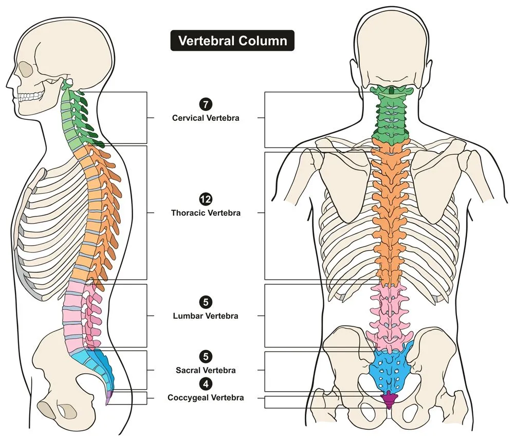
The vertebral column is formed of four different types of vertebrae: the cervical vertebrae, the thoracic vertebrae, the lumbar vertebrae, and the sacrococcygeal vertebrae, in order from head to hip.
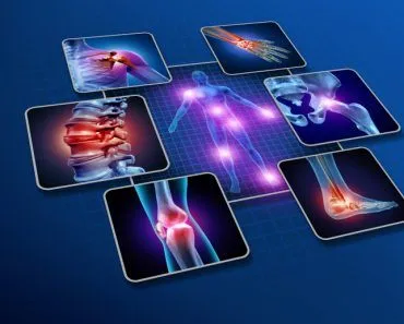
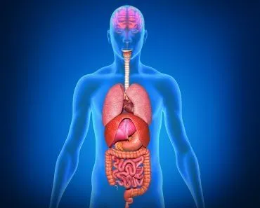
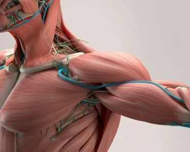
The skeleton is what supports and anchors the soft tissue of our body, such as muscles and skin. It gives mobility and a definite shape, as well as protecting us from environmental stressors. For example, the skull protects the brain, while the ribs protect the lungs and the heart.
Recommended Video for you:
The Skeletal System
It should be obvious that the skeleton is one of the most important parts of our body. The skeleton is made up of bones – tissue composed of structural proteins, such as collagen and minerals, primarily calcium, as well as various bone cells and blood vessels.
There are 206 bones that form our skeleton. These 206 bones can be divided into the axial and appendicular skeleton. The axial skeleton is comprised of the bones that make up our face, neck and backbone (the vertebrae). You can remember this by recollecting that the word axial means a line (imaginary or real) at the center of an object. All bones in the axial skeleton are those which the central axis of the body could pass through.
The appendicular skeleton is formed of all the bones other than the axial bones, such as the bones that form the arms and legs. The word “appendicular” means “limbs”.
In this article, we will look at a particular set of bones within the axial skeleton – the vertebral column. This is the column through which our spinal cord passes—an essential aspect of our existence!
There are 4 different types of vertebrae:
- Cervical vertebrae
- Thoracic vertebrae
- Lumbar vertebrae
- Sacrococcygeal vertebrae
Vertebral Column
Before we delve into the different types of vertebral bones, it’s important to understand the structure and features of all vertebral bones.
The vertebral column picks up from where the skull ends and goes right down to the lower back. It is composed of 31 individual bones called vertebrae, and the entire column is divided into 5 sections – cervical, thoracic, lumbar, sacrum and coccyx – in descending order of location. Each section has a different number of vertebrae, interspersed with intervertebral discs. These discs allow for movement, act as shock absorbers, form fibrocartilaginous joints, etc. They are an important part of the vertebral column.
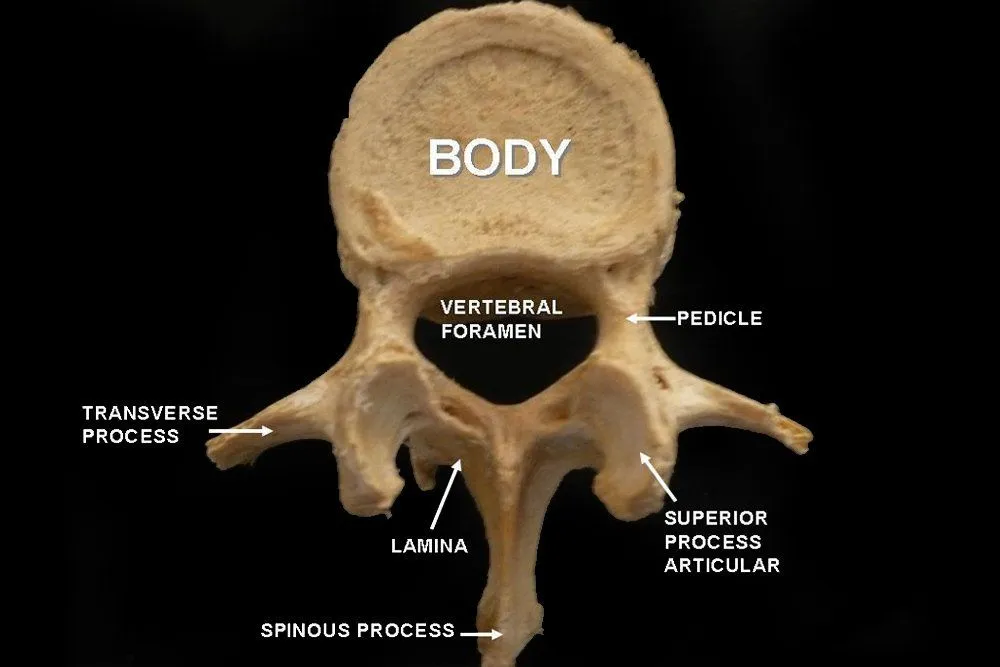
The typical structure of a vertebra is illustrated below:
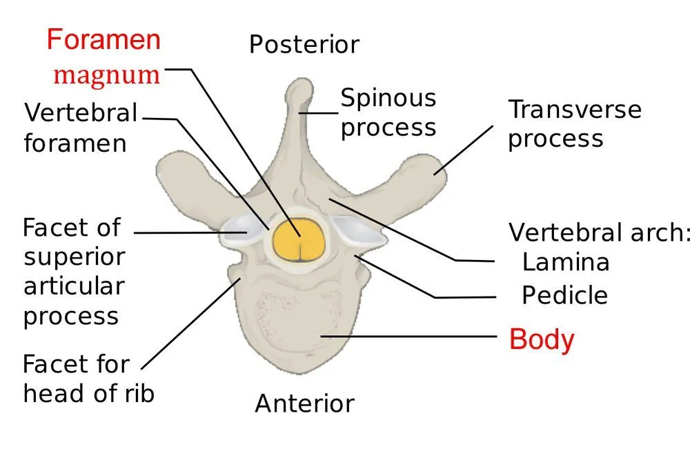
There is a part of the body, the vertebral foramen, where the spinal cord, the process that handles nerves from the brain, passes through. Several other processes exist in the vertebral bone, such as the spinous process, seen in the picture above. These processes are elongated, bony protrusions that help in association and articulation with muscles and other bones.
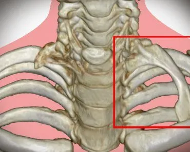
Cervical Vertebrae
These are the bones that form the neck, making up the first section of the vertebral column. They comprise 7 bones – C1 to C7 (this is the naming convention for vertebral bones).
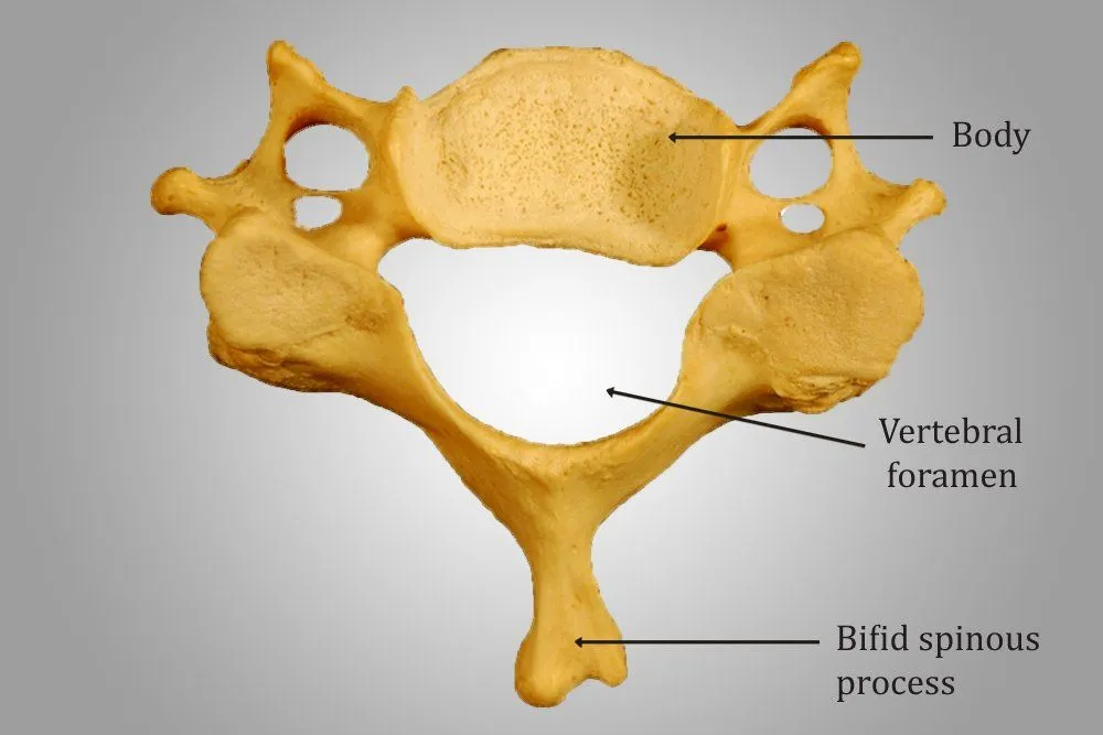
They generally have a smaller body and larger vertebral foramen, and are known as the smallest types vertebrae. Their spinous process is bifid, meaning that the spinous processes are divided into two segments. In the cervical vertebrae, the two segments are unequal.
While the structure of C3 to C6 is similar, C1, C2 and C7 are slightly different, as they are modified for specific functions.
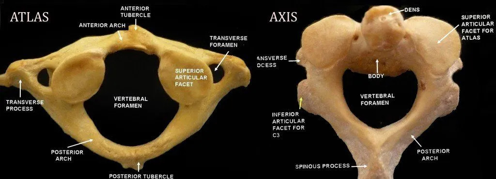
C1 is called the atlas. It has this name because it bears the skull, considered the “globe” of the body. The atlas lacks a body and bears a ring-like structure. It has a wide vertebral foramen to accommodate the thick part of the spinal cord.
Below this is the C2, called the axis. This acts as a pivot upon which the atlas rotates. The most notable part of this bone is the odontoid process, which rises perpendicularly. It is this part that breaks during a ‘judicial hanging’, hitting the medulla and causing death.
If you touch the nape of your neck, a bony protrusion can be felt. This is the C7, also called the vertebra prominens. The protrusion is the spinous process, which is not bifid, of C7.
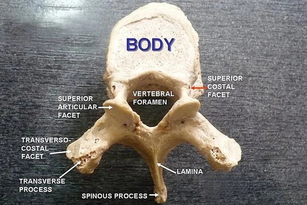
Thoracic Vertebrae
There are 12 thoracic vertebral bones, referred to as T1 – T12.
When you think of the thoracic region, the first thing that comes to mind is the rib cage. The rib cage lies in the thoracic region of the body, and the thoracic vertebrae have a slight modification in order to articulate with the ribs. This modification comes in the forms of demifacets. Facets are basically flattened surfaces of a bone. A demifacet is half of a facet, and is meant for articulation with the ribs and coastal cartilage. The vertebrae have both superior and inferior demifacets. Each rib articulates with the corresponding vertebra and the vertebra above it.
Lumbar Vertebrae
There are five lumbar vertebrae, designated as L1 – L5.
These are the largest vertebrae and have huge bodies. They bear the weight of the body and have a great capacity for movement. The orientation of their process is parasagittal (adjacent to the sagittal or longitudinal plane), which is supposedly responsible for their bending capacity. Spondylosis is most common is this region of the vertebral column.
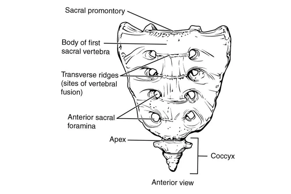
Sacrococcygeal Vertebrae
More often than not, the sacrum and coccyx are studied together as the sacrococcygeal region. The sacrum consists of 5 bones that fuse together to form a single bone, while the coccyx consists of 3-5 bones that typically do the same. In females, the sacrum is short and wide, whereas in males, it is long and narrow. The coccyx is the vestigial remnant of the tail.
Damage to the vertebral column can become extremely dangerous, as it houses the spinal cord. Therefore, one must be careful of even tiny things that affect your spine, such as your position while sitting, lifting weights, etc. Incorrect posture or strain while exercising might put pressure on certain nerves within the spinal cord, not only causing pain, but even leading to far-reaching and disastrous complications.
Life would not be the same—in fact, it would be impossible—without a protected spinal cord working to send signals back and forth from the body and the brain. In other words, do whatever you can to keep it safe!