Table of Contents (click to expand)
The orbicularis oculi is the broad, flat muscle that encircles each eye and is the only muscle capable of closing the eyelids. Its name comes from Latin — orbicularis (“small circle”) and oculi (“of the eye”). Anatomists divide it into three parts: an outer orbital part, a palpebral part inside the eyelids (further subdivided into preseptal and pretarsal portions), and a deep lacrimal part (Horner’s muscle) that helps pump tears across the eye.
The orbicularis oculi is the primary muscle responsible for eyelid closure. Like most facial muscles, the orbicularis oculi muscle too has a complex anatomy. Part of the reason can be attributed to the complicated spatial arrangement of the myofibers and their insertion directly into the skin of the face.
When I was first told by my oncologist that I needed to wear corrective glasses, as my vision had become less than perfect, I was heartbroken. I didn’t want to wear glasses at all!
So, the first thing I did after getting my first pair of spectacles was to try and figure out ways to get my vision back to normal, i.e., 20/20. While that exercise didn’t really bear the intended fruit, I certainly learned a great deal about human eyes.
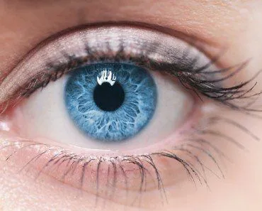
One of the things about eyes that always bothered me, as I’m sure it bothers many of you, is that certain muscles are still working when you close your eyelids to shut them completely.
You see, we’re often told that the best way to relax your eyes is to close your eyes and give them some rest. I, personally, have always associated the idea of ‘resting’ with certain relevant muscles/muscle groups becoming relaxed, which ultimately leads to the relaxation of the concerned body part. Following that logic, one would assume that when the eyelids are closed, no eye muscles are being engaged, because if they were, then those particular muscles wouldn’t really get a chance to relax, right?
That quandary brings us to the question – what happens in the ‘background’ when you close your eyes (more specifically, when you shut your eyelids)?

Let me introduce the main muscle behind all this…
Recommended Video for you:
Orbicularis Oculi
The orbicularis oculi is the primary muscle responsible for eyelid closure. The name itself tells you what it does and where it sits: orbicularis comes from the Latin orbiculus, meaning a small disk or little circle, and oculi is the Latin genitive of oculus — “of the eye.” Put together, it is literally the “little circular muscle of the eye,” a flat sphincter-like band of fibers that encircles the orbital opening (Source).
Like most facial muscles, it has a complex anatomy. Part of the reason can be attributed to the complicated spatial arrangement of the myofibers and their insertion directly into the skin of the face, which is what lets the muscle do everything from a soft, unconscious blink to a tight, forceful squeeze.
The orbicularis oculi is not only responsible for actively closing the eyes, but also for much of the facial expression made around the eyes — squinting, wincing, and the wrinkling that produces “crow’s feet” at the outer corners. Anatomists divide the muscle into three distinct parts: the orbital part (the outer ring overlying the bony orbital rim), the palpebral part within the eyelids (further subdivided into preseptal and pretarsal portions), and the deep lacrimal part, also known as Horner’s muscle (Source).
Orbicularis Oculi Structure And Anatomy
The orbital portion of the muscle has a reddish color and forms a complete ellipse overlying the orbital rim and the cheek. It originates from the medial palpebral ligament, the nasal part of the frontal bone, and the frontal process of the maxilla, and its fibers loop laterally around the eye without interruption. This is the thickest part of the muscle and is mainly responsible for the forceful, voluntary eyelid closure you use when squinting into bright sunlight or screwing the eyes tightly shut.
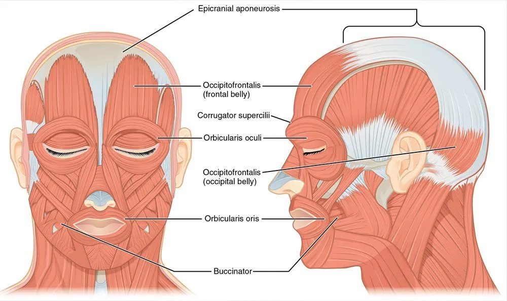
The palpebral portion sits within the eyelids themselves and is responsible for the gentle, involuntary blink. It is conventionally subdivided into two layers based on what they overlie: the preseptal portion, which is thin and pale and is formed by two half-ellipses of muscle fibers bifurcated by the medial and lateral canthal tendons, and the pretarsal portion, which lies directly anterior to the upper and lower tarsal plates and produces the rapid blink reflex.
The deepest slip of the muscle is the lacrimal portion, also called Horner’s muscle, after the American anatomist William E. Horner who described it in 1824. It arises from the posterior crest of the lacrimal bone, passes behind the lacrimal sac, and inserts into the medial ends of the eyelids. Each time you blink, this small muscle squeezes the lacrimal sac and tugs the puncta inward, helping to pump tears off the surface of the eye and down the nasolacrimal duct (Source).
Innervation And Blood Supply
The orbicularis oculi is supplied by the facial nerve (cranial nerve VII). Its temporal branch innervates the upper half of the muscle along with the frontalis and corrugator supercilii, while the zygomatic branches supply the lower half. Sensation around the eye comes from branches of the trigeminal nerve, but the order to contract the muscle and shut the lid travels exclusively along the facial nerve. Blood is supplied by the facial, superficial temporal, and ophthalmic arteries.
Orbicularis Oculi Functions
The main function of the orbicularis oculi muscle is to close the eyelids, and it is in fact the only muscle that can do so. It is so critical that if either the muscle itself or its motor supply — the facial nerve (CN VII) — is injured, the patient loses the ability to fully close that eye, a condition called lagophthalmos. This is the hallmark sign on the affected side of Bell’s palsy, where facial nerve inflammation paralyses the orbicularis oculi and the eye must be lubricated and protected to prevent corneal damage.
The palpebral portion of the muscle acts involuntarily, closing the eyelids gently — most obviously during the spontaneous blink that resurfaces your tear film roughly every five seconds, and in the protective corneal reflex that snaps the lids shut when something approaches the eye.

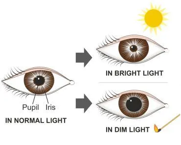
When the entire orbicularis oculi muscle is brought into action, the skin of the forehead, temple, and cheek is drawn toward the medial angle of the orbit, and the eyelids are firmly closed, as occurs in photophobia.
Tear Drainage And The Lacrimal Pump
Beyond shutting the eye, the orbicularis oculi works as a tear pump. Each blink, the contraction of the lacrimal (Horner’s) part draws the puncta and lacrimal canaliculi medially while compressing the lacrimal sac, propelling tear fluid from the eye’s surface down the nasolacrimal duct into the nose. Without that pumping action, tears spill over the lid margin instead of draining — which is why people with weakened orbicularis oculi tone (after a stroke or in older age) often experience watery eyes.
Clinical And Cosmetic Significance
Because of how visible and active this muscle is, it shows up in several familiar medical and cosmetic contexts:
- Bell’s palsy and other facial-nerve injuries paralyse the orbicularis oculi on one side, leaving the eye unable to close fully (lagophthalmos) and at risk of exposure keratitis.
- Benign essential blepharospasm is a focal dystonia in which the orbicularis oculi contracts involuntarily and repeatedly, forcing the eyes shut and disrupting reading, driving, and other daily tasks.
- Botox for crow’s feet works by injecting botulinum toxin into the lateral fibers of the orbicularis oculi to relax them. Crow’s feet are produced by repeated contraction of these fibers, and chemodenervation typically softens those dynamic lines for three to four months. Neuromodulator injections targeting the orbicularis oculi remain one of the most common cosmetic procedures performed worldwide.
- Lower-lid blepharoplasty often involves tightening the orbicularis oculi to address sagging and prolapsed fat as the muscle weakens with age.
Are Some Eye Muscles Still Engaged When You Close Your Eyes?
You actually have different muscles that open and close your eyes. As mentioned earlier, the orbicularis oculi muscle helps to close your eyelid, while the levator palpebrae superioris muscle assists in opening it.
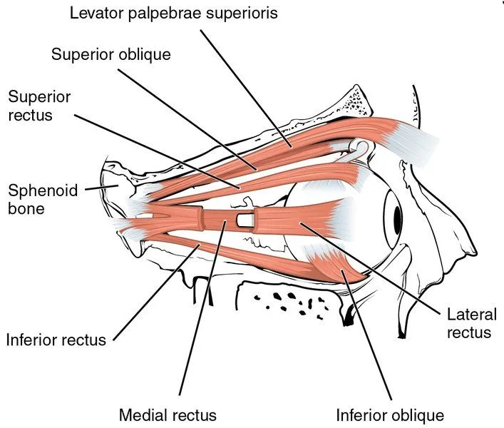
There’s also a third muscle that controls the upper eyelids, but only in special circumstances, such as when you are surprised/shocked. You cannot control this muscle actively; it fires all on its own.
When all these muscles are relaxed, your eyelids will be somewhere in the middle of being closed and opened, quite similar to how it looks when you’re tired!
So, to be fair, at any given moment, your eyelids are in the position they are due to the combined function of these muscles, which work in tandem. This is why it’s fair to say that, yes, even when you close your eyes, some muscles ARE still engaged, meaning that even when you’re sleeping, your body is never fully relaxed!