Physics
Astrophysics
Theoretical Physics
Sports
Super Heroes
Earth Science
Chemistry
Biology
Botany
Zoology
Medicine
Neuroscience
Engineering
Technology
Artificial Intelligence
Computing
Mathematics
Social Science
Psychology
History
Sociology
Geography
Philosophy
Economics
Linguistics
Art
Videos
About Us
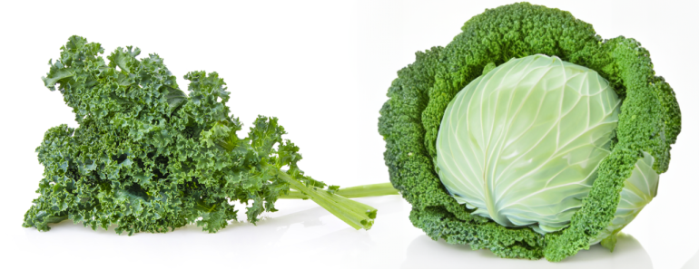
Are Kale And Cabbage The Same Plant?
April 22, 2024
Botany
What Are The Three Waves Of Feminism?
April 17, 2024
Psychology
How Are Memories Formed And Recalled?
April 16, 2024
Psychology
Were Pirates The Revolutionaries Of The High Seas?
April 15, 2024
Sociology
Why Is Shakespeare Still Taught In Schools Today?
April 14, 2024
Economics
Why Does Sleep Deprivation Cause Body Aches?
April 12, 2024
Psychology
Why Were Lawless Pirates Bound By Codes Of Conduct?
April 8, 2024
Sociology
Why Are Insects Attracted To Artificial Lights?
April 3, 2024
Zoology
Why Are There More Men In The World Than Women?
April 2, 2024
Economics
How Is Einstein’s Theory Of Relativity Related To GPS?
April 1, 2024
Physics
Can High Heat Really Fry Your Electronics?
March 25, 2024
Technology
Previous