Table of Contents (click to expand)
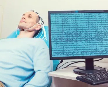
An ultrasound machine uses sound waves to create images of the inside of the human body. A transducer probe sends out sound waves, which bounce off of fluids, tissues, and organs and are received by the probe. The computer uses this information to create a real-time image of the inside of the body.
Back when medical technology was not as developed as it is today, physicians had to ‘feel’ the affected organ of their patients’ bodies and then use their experience to determine the underlying cause of the discomfort.
Having no other form of examination at their disposal, our ancient ancestors had to depend entirely on this crude, rudimentary technique of detecting problems inside the body. Fast forward to today, and we can spot the smallest malignant growth anywhere inside the human body, thanks to ultrasound imaging.
Recommended Video for you:
What Is Ultrasound Scanning?
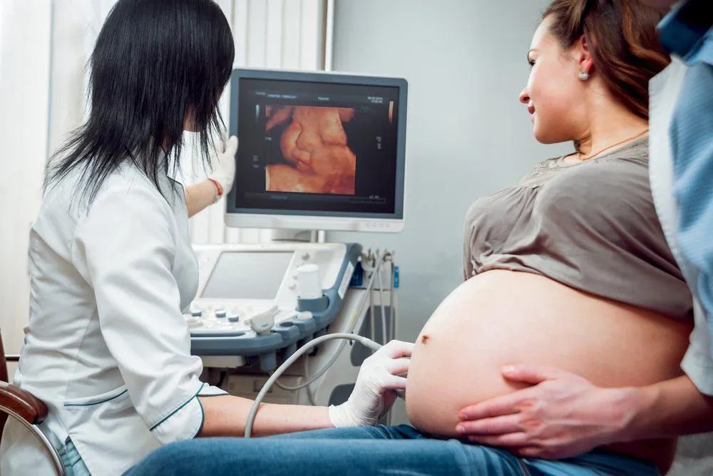
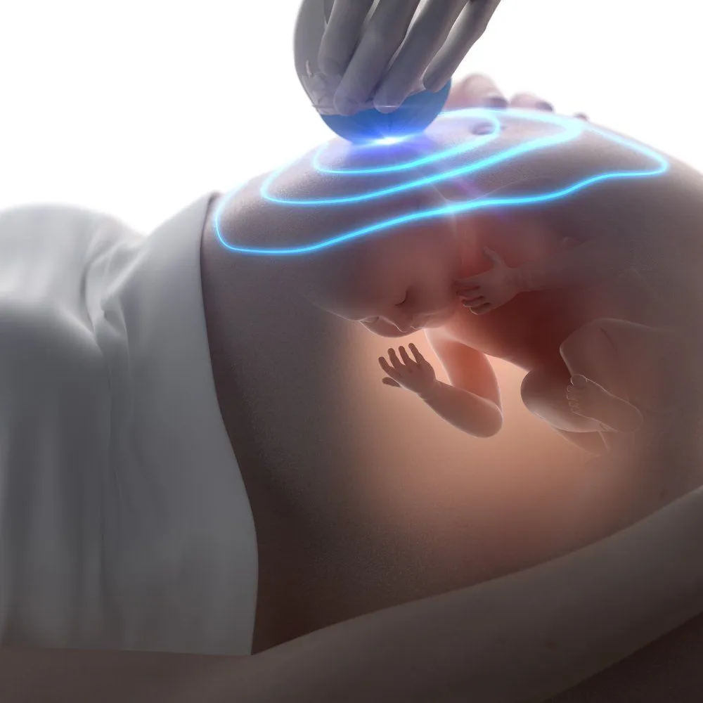
Ultrasound scanning, or simply ultrasound, is a non-invasive and painless diagnostic technique used to “look inside” the innards of a human body. Also known as sonography, it’s quite an effective and widely used technique that doctors and medical experts use to determine what’s happening inside the human body without actually operating on it. Although it can be (and is) used in nearly all cases where the state of a patient’s internal organs must be determined (such as to check blood circulation in a limb, detection of tumors etc.), sonography is most often associated with pregnancy, as it helps to determine various parameters of fetal development, which helps to calculate the due date.
The Ultrasound Machine
Unlike an X-ray machine, which uses electromagnetic waves to form images of the human body, ultrasound machines use sound; more specifically, it uses ultrasound waves (hence the name ‘ultrasound machine’) to find out what’s going on inside the body.
Components
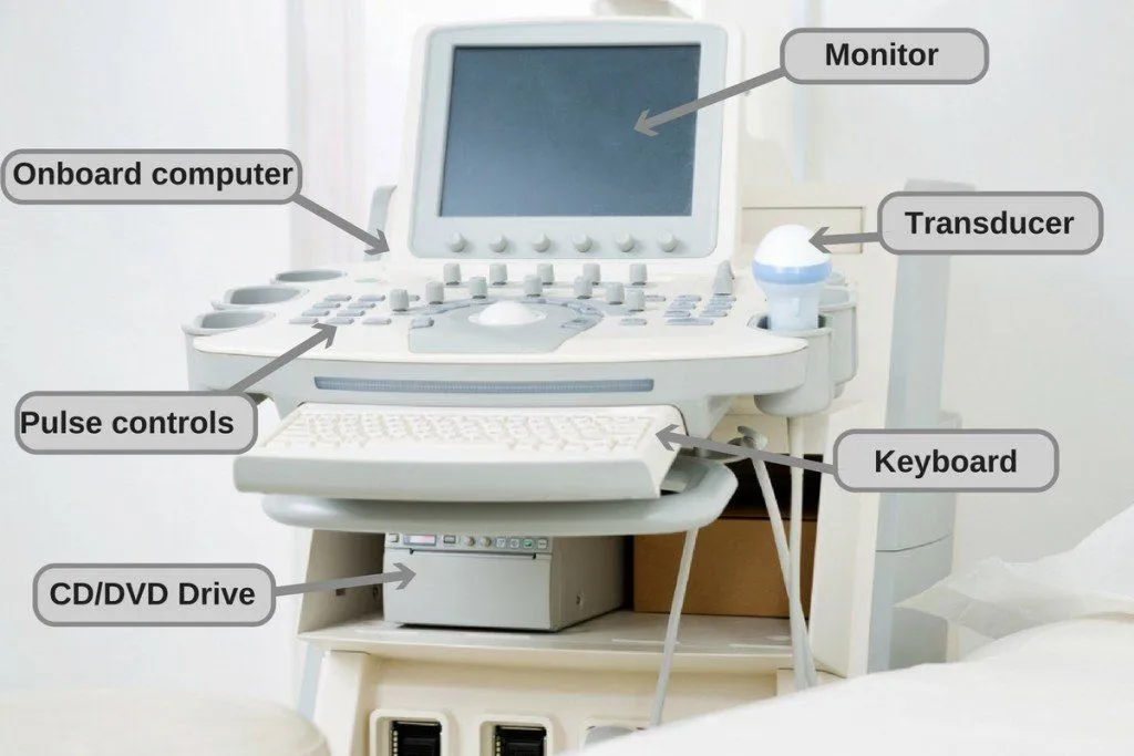
Structurally, an ultrasound machine is very simple. It consists of two basic parts:
1. A transducer probe that sends sound waves and receives their echoes
2. A computer (a central processing unit or CPU) that supplies power to the transducer probe, performs all the calculations and shows the images of the internal area on a screen.
How Does Sonography Work?

The transducer probe comes in various shapes and sizes. Irrespective of their appearance, most of them contain one or more piezoelectric elements. Early transducers used natural quartz, but virtually every modern medical probe uses a synthetic ceramic such as lead zirconate titanate (PZT) or, in higher-end systems, single-crystal piezoelectrics, which produce stronger signals across a wider bandwidth. The interesting thing about these materials is that they change shape, or vibrate, when an electric current is applied to them. So, when the transducer is switched on, the elements create vibrations that produce sound waves with frequencies (typically between 2 and 20 MHz for diagnostic imaging, and as high as 30–70 MHz for ultra-high-frequency probes) that are far higher than what humans can hear.
Before starting the process, a water-based gel is applied directly to the skin on the designated area of the subject’s body. The gel displaces air between the probe and the skin — and this matters more than it sounds. Air has an acoustic impedance roughly 3,500 times lower than soft tissue, so even a thin air gap reflects more than 99.9% of the sound waves back at the probe before they ever reach the body. The gel’s impedance closely matches that of skin, allowing sound to pass through almost without loss. The sound beam produced from the probe is focussed either by the shape of the probe itself, a lens in front of the probe, or a set of control pulses from the scanner, resulting in an arc-shaped sound wave emanating from the face of the probe, entering the body and finally coming into focus at the desired depth.
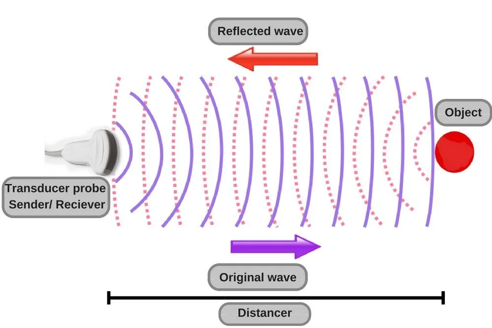
The transmitted sound waves pass through the thin layer of skin, but bounce off fluids, tissues and internal organs wherever there is a change in acoustic impedance. These reflected waves are received by the same probe — the piezoelectric effect runs both ways, so the returning echoes squeeze the elements and generate a tiny voltage that is fed into the computer. The computer uses the speed of sound in tissue (roughly 1,540 metres per second) and the time taken by each echo to return to work out how deep the reflecting structure lies, and the strength of the echo determines how bright that point appears on screen. Stitch enough of these points together and you get a real-time image of the inside of the human body.
Traditional ultrasound scanners display 2-dimensional images, meaning that images are displayed in flat sections of the body. However, modern scanners use the same calculations to assemble 3-dimensional volumes, and 4D systems update those volumes many times per second to show movement in real time — a wonderful advancement, especially for would-be parents watching the growth of their baby. The technology has also shrunk dramatically: handheld point-of-care probes such as the Butterfly iQ+, GE Vscan Air and Mindray TE Air now plug into a smartphone or tablet and deliver image quality that was confined to cart-sized machines a decade ago. AI is the other big shift — recent systems use deep-learning models to auto-measure fetal biometry, flag suspected anomalies on prenatal scans, and clean up images in real time so less-experienced operators can still capture diagnostic-quality views.
There are a few different types of ultrasonography, depending on the type of examination required, including Doppler ultrasonography (to determine whether certain structures are moving away from or towards the probe), contrast ultrasonography (used in echocardiography and for lesion characterization), and molecular ultrasonography.
If not for this simple, yet incredibly effective machine, not only would we be unable to locate a tumor or diagnose a disease inside the human body, but we would be deprived of the heartwarming sight of watching a new life grow inside the womb.